
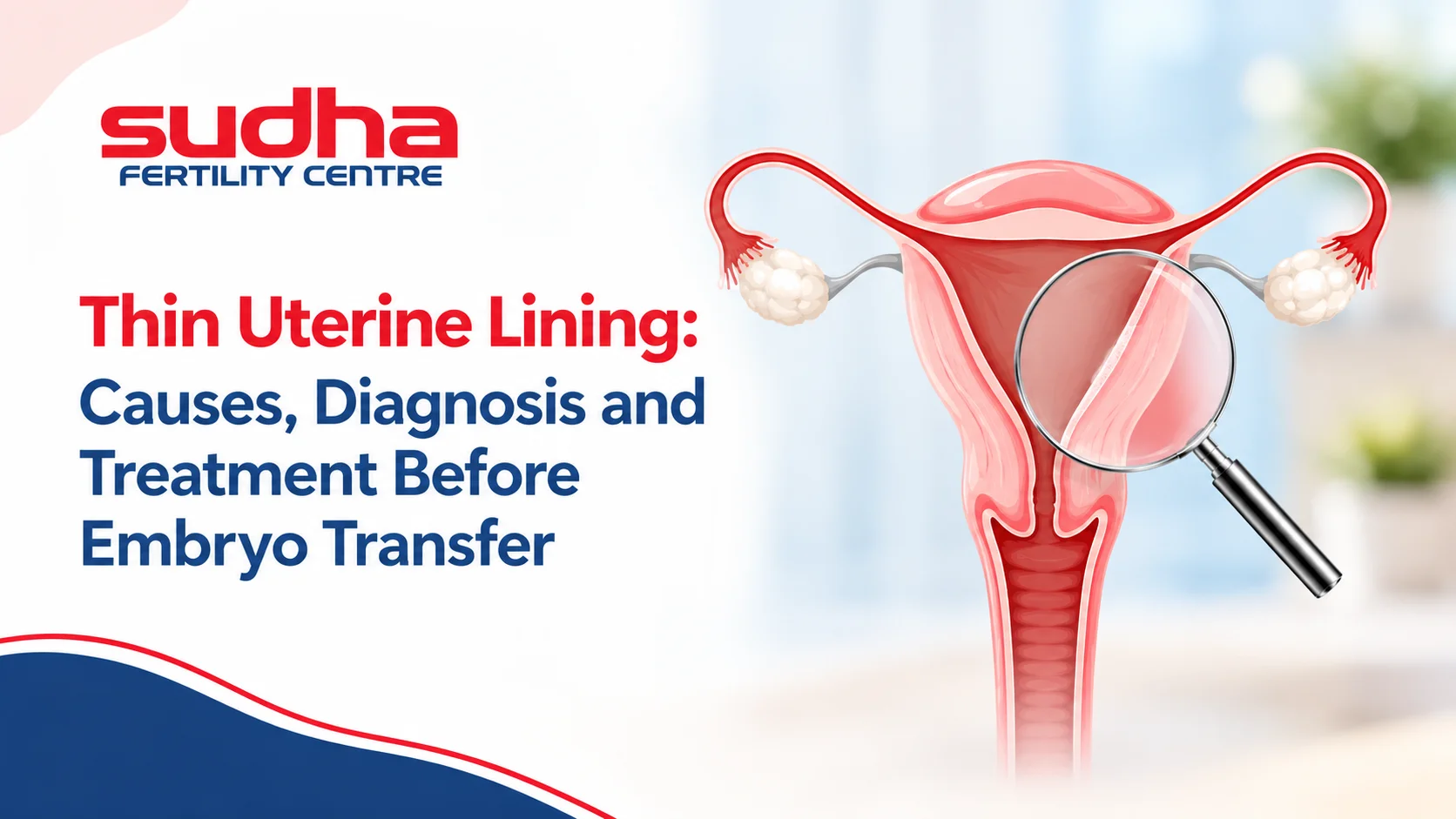
Thin Uterine Lining: Causes, Diagnosis and Treatment Before Embryo Transfer
Medically Reviewed
Written By: Dr. S. Pradeepa Sudhakar, MBBS, DGO | Senior Fertility Specialist & Vice President, Sudha Fertility Centre
Medically Reviewed By: Dr. S. Dhanabagyam, MBBS, DGO | Founder-President & Senior Consultant, Sudha Fertility Centre
About the Authors
- Dr. S. Pradeepa Sudhakar is a Senior Fertility Specialist with over 15 years of clinical experience. An alumna of Madras Medical College, she specializes in managing complex reproductive profiles, customised ovulation induction, and advanced follicular tracking. As Vice President, she focuses on providing clear, evidence-based guidance to couples starting their families.
- Dr. S. Dhanabagyam is the Founder-President of Sudha Fertility Centre and a reproductive medicine pioneer who has guided our clinical teams in helping bring over 100,000 (1 lakh) healthy babies into the world. With over 40 years of clinical excellence, she peer-reviews our patient guides to ensure medical accuracy, transparency, and clinical integrity.
Quick Summary
- The Clinical Timeline: Evaluating your requirements for a thin uterine lining treatment protocol happens dynamically through tracking scans right before your progesterone exposure begins.
- The Biological Causes: Inadequate tissue thickness is driven by factors like low oestrogen absorption, intrauterine scarring, chronic endometritis, or underlying pelvic circulatory challenges.
- The Diagnostic Connection: Specialists utilize high-resolution transvaginal scans and diagnostic hysteroscopy to verify the structural health and vascular patterns of your inner womb tissue.
- The Clinical Precautions: Certain adjuvant medical therapies are classified as off-label or investigational, requiring clear clinical transparency regarding their current evidence boundaries.
- The Next Treatment Steps: Transitioning from a fresh cycle to a rested frozen embryo transfer allows your care team to prepare your tissue utilizing physiological hormone protocols.
Thin Uterine Lining: Causes, Diagnosis and Treatment Before Embryo Transfer
When a routine ultrasound scan reveals that your womb lining is too thin, it can feel like a sudden adjustment to your pregnancy plans. Many couples assume that securing a high-grade embryo is the only major challenge, but that embryo cannot stick without a thick, healthy tissue bed to protect it. Fortunately, entering a targeted thin uterine lining treatment protocol helps rebuild your inner tissue layers by addressing hormone absorption or local blood circulation.
This patient guide simplifies the biological causes behind an inadequate womb lining, translates diagnostic checkmarks, and details the standard and investigational options used to prepare your body for a successful transfer.
The Biological Threshold: What Defines a Thin Lining?
Your uterine lining is known clinically as the endometrium. This is the specialized inner tissue layer where a fertilized embryo must stick to establish a pregnancy. Throughout a normal menstrual cycle, your body naturally releases oestrogen. This hormone encourages this tissue layer to multiply, grow thick, and build a dense network of blood vessels.
In the world of assisted reproduction, fertility specialists utilize a biological threshold to measure endometrial readiness. Many clinics use 7 millimetres as a working thickness threshold on an ultrasound scan right before your progesterone exposure begins. Furthermore, the tissue should display a distinct, structural pattern under the scan known as a triple-line configuration. The triple-line design signals that the tissue is fully organized and receptive. If your tracking measurements stay persistently below 7 millimetres, the tissue is classified as a thin endometrium, which requires focused medical review before an embryo can be safely transferred.
Why Inadequate Endometrial Thickness Occurs: The Common Causes
When your lining fails to grow adequately, an underlying structural or hormonal challenge is usually running quietly behind the scenes. Doctors evaluate several primary biological factors before mapping out a protocol:
1. Inadequate Oestrogen Levels or Poor Cellular Absorption
Oestrogen is the primary fuel your body uses to build up the womb lining during the first half of your cycle. Sometimes your ovaries struggle to produce enough natural oestrogen. Alternatively, the receptors inside your uterine tissue may be sluggish. When receptors fail to absorb the hormone patches or pills you are prescribed, the lining remains flat and unsupportive.
2. Physical Scarring and Womb Cavity Trauma
Past surgical procedures inside the uterus can leave behind physical changes that prevent healthy tissue growth. A primary example is Asherman syndrome. This condition happens when past surgeries like a dilation and curettage (D&C) or cavity-entering removals for large Uterine Fibroids create bands of internal scar tissue. These tough scar bands replace your healthy endometrial cells, making portions of the lining blind to oestrogen signals.
3. Chronic Tissue Inflammation and Genital Tuberculosis
Underlying infections inside the womb can severely stunt tissue growth. For instance, chronic endometritis causes a persistent, low-grade bacterial inflammation of the inner lining. This inflammation directly alters your cellular receptivity and halts normal tissue expansion.
These severe pelvic infections frequently track upward through the reproductive tract. This destructive migration can simultaneously create internal uterine scarring and lead to Blocked Fallopian Tubes. Furthermore, in India, endometrial health challenges are frequently driven by genital tuberculosis. This specific infection silently scars the endometrial bed, leading to a thin, non-responsive lining.
4. Compromised Pelvic Blood Circulation and Medication Blocks
Your uterine lining cannot multiply without a continuous supply of oxygen-rich blood. General circulatory health factors can contribute to local vascular changes inside the reproductive tract. Furthermore, women who take specific ovulation medications like clomiphene citrate for multiple consecutive cycles can experience a temporary thinning of the lining. This medication can block oestrogen receptors inside the uterus, creating a temporary drop in thickness that requires a change in your medication protocol.
How a Thin Uterine Lining Is Diagnosed
To build a secure treatment plan, your fertility team must look beyond a single thickness measurement. They need to analyze the actual health, structural consistency, and circulation patterns of your tissue. This diagnostic roadmap consists of three core evaluations:
- Serial Transvaginal Ultrasound Tracking: High-resolution scans measure your lining thickness dynamically through the cycle. Specialized color Doppler imaging maps the blood flow inside the uterine arteries to check if the tissue is receiving an adequate blood supply.
- High-Precision Diagnostic Hysteroscopy: Your consultant guides a tiny camera gently through your cervix to view the entire inside of your womb cavity. This allows them to physically spot and clear away hidden Asherman Syndrome scar tissue bands, small polyps, or micro-inflammation that an ultrasound scan might miss.
- Endometrial Receptivity Tracking: If you face repeated transfer delays despite having a lining that looks structurally normal, your team may look into a specialized biopsy cycle. While global trial evidence has been mixed and routine use is not recommended for every patient, this tool is reserved for selected cases to analyze the genetic timing window of your tissue.
Treatment Options for a Thin Uterine Lining
Discovering that your lining is thin does not mean you have run out of choices. Our clinical teams utilize a multi-step medical toolkit to encourage your endometrial cells to expand safely. It is vital to separate standard, evidence-backed care from adjuvant, investigational options:
Standard First-Line Medical Therapies
- Maximizing and Diversifying Oestrogen Delivery: If standard oral oestrogen pills fail to grow your tissue, your doctor will bypass your digestive system to improve first-pass hepatic metabolism. They will pivot your protocol to include high-purity transdermal oestrogen patches applied directly to your skin, or prescribe localized vaginal oestrogen inserts to deliver the hormone straight to your tissue receptors.
- Hysteroscopic Adhesiolysis for Scar Removal: If your diagnostic hysteroscopy reveals that your womb cavity is being compressed by internal scar tissue, your surgeon can use micro-scissors during the same procedure to safely clear away the scar tissue, freeing your underlying endometrial layer to grow normally again.
Evidence-Limited and Investigational Adjuvant Therapies
- Oral Pentoxifylline Treatment: This medication is used off-label and selectively in small studies, often combined with vitamin E, to help improve local vascular patterns. While it is used to support general circulatory paths, clinical trial data regarding its standalone efficacy for endometrial thickening remains limited.
- Vaginal Sildenafil Suppositories: This is an investigational, off-label compounded preparation used selectively to dilate pelvic blood vessels. It is not universally available, must be custom-compounded, and carries potential side effects like headaches or flushing. Clinical data do not support the routine addition of low-dose oral aspirin for this specific purpose, as routine aspirin use fails to demonstrate a significant increase in live birth rates or thickness metrics in large clinical sets.
- Uterine Platelet-Rich Plasma (PRP) Infusion: This is an experimental, investigational therapy reserved for stubborn lining profiles. Your medical team draws a sample of your blood and spins it in a laboratory centrifuge to isolate your natural growth factors. Your specialist infuses this plasma gently into your uterine cavity using a soft catheter a few days before your scheduled transfer. Because this catheter procedure carries minimal but recognized risks of localized infection, bleeding, or venepuncture discomfort, it should be offered within a clearly consented framework. It is an optional adjuvant and does not guarantee a successful thickening outcome.
Strategic Protocol Adjustments: Fresh vs Frozen Transfer
When your lining thickness stays borderline during an active treatment cycle, your choice of transfer timing becomes a powerful tool to protect your biological outcomes. Your care team will look closely at your records to help you choose the best strategic path:
- The Fresh Transfer Limitation: During a fresh IVF cycle, your ovaries are heavily swollen from high-dose egg-retrieval stimulation medications. This creates an intensely high, artificial oestrogen environment in your bloodstream. This shift can cause your uterine lining to mature too quickly, creating a structural mismatch that makes it harder for an embryo to stick safely.
- The Frozen Embryo Transfer Advantage: If your lining stays thin during a fresh cycle, your specialist will safely freeze your healthy blastocysts and halt the cycle. Shifting your timeline toward a specialized Fresh vs Frozen Embryo Transfer pathway allows your body to clear out those heavy stimulation drugs completely. Your doctor can then prepare your lining during a future month using gentle, physiological hormone doses that focus entirely on building thickness inside a fully rested body during a dedicated Frozen Embryo Transfer (FET) cycle.
Frequently Asked Questions
Can lifestyle changes or specific diets completely cure a thin uterine lining?
What is the absolute minimum lining thickness needed to safely proceed with a transfer?
How does a clinical uterine PRP therapy help improve my lining thickness?
Will having a thin lining cause a permanent risk of a future miscarriage?
Can a hysteroscopy checkup fix a thin lining during the very same procedure?
Ready to Align Your Body for Conception?
A thin uterine lining report is a manageable clinical checkpoint on your road to building a family, and starting a targeted thin uterine lining treatment protocol allows you to prepare your body step by step. Finding a borderline thickness measurement simply means your womb requires a customized, data-driven approach to unlock its reproductive potential before your transfer cycle begins. At Sudha Fertility Centre, our experienced medical teams focus on providing advanced laboratory infrastructure, transparent evaluations, and compassionate care to help you review your options.
Our clinical teams will review your baseline results with you and set out the precise options available in your case. Contact us today to schedule your complete evaluation at our nearest branch location.
Disclaimer: The information provided in this article is for educational purposes only and does not substitute for professional medical advice, diagnosis, or treatment regarding your uterine health. Always consult a qualified physician, gynaecologist, or certified fertility specialist to discuss your unique health concerns before making any sudden changes to your daily routine, lifestyle habits, or medical protocols.

Dr. S. Pradeepa is a fertility specialist at Sudha Fertility Centre, Erode, with expertise in IVF, IUI, ICSI, PCOS, and endometriosis. She holds MBBS, DGO, DNB (OG), and a Fellowship in Reproductive Medicine. Known for her patient-centric approach, she provides personalized, evidence-based care and reviews medical content to guide informed fertility decisions.