

Blocked Fallopian Tubes: Causes, Symptoms, Diagnosis and Treatment Options
Types of Fallopian Tube Blockage: Location, Causes & Treatment
-
- Proximal Blockage – occurs at the uterotubal junction near the uterus; often caused by tubal spasm, PID scar tissue, or endometriosis; treated effectively with tubal cannulation after laparoscopy confirmation
- Mid-Segment Blockage – affects the middle portion of the tube; commonly linked to ectopic pregnancy history, tubal ligation reversal, or surgical scarring; IVF is often preferred due to high recurrence risk after surgery
- Distal Blockage (Mild) – located near the ovary with partially damaged fimbriae; caused by mild endometriosis or previous infection; laparoscopic fimbrioplasty or salpingostomy offers a 27–60% pregnancy rate with ~10% ectopic risk
- Distal Blockage (Hydrosalpinx) – near the ovary with the tube distended and filled with fluid; caused by severe PID, advanced endometriosis, or previous infection; salpingectomy before IVF is ASRM-recommended as untreated hydrosalpinx reduces IVF success by approximately 50%
The location of the blockage not just its presence determines which treatment gives the best chance of pregnancy.
Receiving an HSG result that shows a blocked fallopian tube can feel like the ground has shifted. A moment ago the plan was straightforward; now there is a diagnosis, unfamiliar medical terms, and a lot of unanswered questions about what this means for the chances of getting pregnant.
Blocked fallopian tubes are one of the most common causes of female infertility, accounting for 25 to 40% of cases. But they are also one of the most treatable, with clear diagnostic and treatment pathways that depend on where the blockage is, what caused it, and what the overall fertility picture looks like. This guide covers all of that, including the one piece of information most articles skip over entirely: why an untreated blocked tube can significantly reduce IVF success rates, and what to do about it.
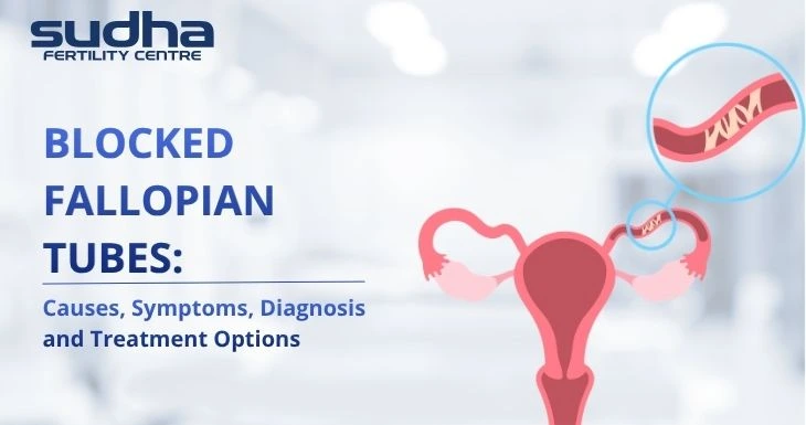
What Do Fallopian Tubes Actually Do?
After ovulation, the egg is captured by the fimbriae, the finger-like projections at the end of the fallopian tube, and drawn into the tube. Sperm travel from the uterus up into the tubes, where fertilization takes place. The fertilized egg then travels down the tube over the next three to four days to reach the uterus, where implantation occurs.
The tube is not a passive channel. It is lined with tiny hair-like cells called cilia that actively move the egg and embryo toward the uterus through coordinated wave-like motion. The tube also provides the correct environment for fertilization and early embryo development. When the tubes are blocked or damaged, any step in this process is disrupted.
This is why even partial repair of a damaged tube does not always restore full function. Surgery can open a blocked tube, but it cannot always restore the cilia or the internal environment that the tube needs to function properly. This distinction matters when weighing surgery against IVF.
Types of Fallopian Tube Blockage
Where the blockage is located has a significant impact on what treatment is appropriate and what the chances of success are. There are four main categories.
|
Type |
Location | Common Causes |
Treatment Approach |
| Proximal | Near the uterus (uterotubal junction) | Tubal spasm (often false positive on HSG), scar tissue from PID, endometriosis | Tubal cannulation works well. Always confirm with laparoscopy before surgery. |
| Mid-segment | Middle of the tube | Previous ectopic pregnancy, prior tubal ligation reversal, surgical scarring | Surgical repair is more complex. IVF often preferred as recurrence risk is high |
| Distal (mild) | Near the ovary; fimbriae partially damaged | Mild endometriosis, previous infection | Laparoscopic fimbrioplasty or salpingostomy in young women; 27 to 60% pregnancy rate; ectopic risk around 10% |
| Distal (hydrosalpinx) | Near the ovary; tube distended with fluid | Severe PID, advanced endometriosis, previous infection | Salpingectomy before IVF is ASRM-recommended. Fluid from hydrosalpinx reduces IVF success by approximately 50% if untreated |
Causes of Blocked Fallopian Tubes
Understanding the cause matters because it affects both the type of blockage and the treatment options available.
Pelvic inflammatory disease (PID):
This is the most common cause of tubal blockage in India. PID is an infection that spreads from the cervix through the uterus to the fallopian tubes, causing inflammation that leaves behind scar tissue and adhesions. Importantly, PID is frequently silent. Many women have no idea they have had it because it can occur without obvious symptoms, particularly when caused by certain infections. This is why STI screening is important as part of a fertility workup.
Endometriosis:
Around 30 to 50% of women with endometriosis some degree of tubal involvement and Endometrial-like tissue growing on or around the tubes creates adhesions and scar tissue that can narrow or block the tube. Endometriosis is increasingly being diagnosed in South India due to better awareness, and is often discovered for the first time during an infertility investigation.
Previous ectopic pregnancy:
Treatment for an ectopic pregnancy, whether surgical or medical, can leave scar tissue in the affected tube. Women with a history of ectopic pregnancy are at elevated risk of tubal factor infertility and should begin fertility investigations early when trying to conceive again.
Prior pelvic or abdominal surgery: Adhesions (internal scar tissue) from ovarian cyst removal, myomectomy, appendicectomy, or Caesarean section can cause the tubes to kink, twist, or adhere to surrounding structures. C-section rates in urban South India are high, and adhesions from prior Caesarean are an underappreciated tubal risk factor.
Uterine fibroids: Large fibroids can press on or grow into the cornual junction where the tube meets the uterus, causing proximal blockage. Fibroids are common in reproductive-age Indian women and should always be assessed alongside tubal patency.
Sexually transmitted infections: Chlamydia and Gonorrhoea specifically damage the ciliated lining of the fallopian tube, even when asymptomatic. Repeated infections multiply the damage. STI screening is underperformed in Indian fertility workups and should be included in the initial evaluation.
Symptoms: Why Most Women Do Not Know
Blocked fallopian tubes are almost always silent. There is no pain, no unusual discharge, no warning sign. The most common way the diagnosis is made is through a fertility investigation after a woman has been unable to conceive.
In some specific situations, there may be associated symptoms:
- Hydrosalpinx a fluid-filled blocked tube, can sometimes cause a persistent dull ache or feeling of fullness on one side of the lower abdomen
- Endometriosis-related blockage may be associated with painful periods, pelvic pain, or pain during intercourse, though these symptoms relate to the endometriosis itself, not the tubal blockage directly
- PID-related blockage may follow a history of pelvic pain and fever, though many women with PID-related tubal damage never experienced obvious symptoms at the time
The key message: the absence of symptoms does not mean the tubes are open. Asymptomatic tubal damage is the rule, not the exception. The only way to know is through a diagnostic test.
How Are Blocked Tubes Diagnosed?
Three main tests are used, and they have different strengths and limitations.
HSG (Hysterosalpingography): This is the standard first-line test. Contrast dye is injected through the cervix into the uterus, and an X-ray shows whether the dye flows freely through the tubes and spills into the pelvic cavity. It is a fast, outpatient procedure done between days 7 and 10 of the menstrual cycle. HSG is reliable in most cases, but there is a known 15 to 20% false-positive rate for proximal blockage, often caused by tubal spasm during the procedure rather than a true block. For this reason, a proximal blockage finding on HSG should always be confirmed with laparoscopy before any surgical treatment is planned.
Sonosalpingography (SSG) or Hysterosonography: This uses saline and ultrasound instead of dye and X-ray, with no radiation exposure. It provides good assessment of uterine structure alongside tubal patency and is a useful alternative or complement to HSG, particularly in women who cannot undergo the X-ray procedure.
Laparoscopy: Laparoscopy is the gold standard for diagnosing tubal blockage. Direct visualisation of the tubes and pelvis allows the doctor to assess the severity and exact location of the blockage, identify adhesions or endometriosis, and in many cases treat the problem in the same procedure. Laparoscopy is more invasive than HSG but provides the definitive diagnosis and can be both diagnostic and therapeutic in one step.
Transvaginal ultrasound can sometimes detect hydrosalpinx when the tube is significantly distended, but it misses many cases of tubal blockage. It is not a reliable standalone test for tubal patency.
Treatment Options: The Surgery vs IVF Decision
The right treatment depends on where the blockage is, how severe the damage is, the woman’s age, whether there are other fertility factors, and how quickly conception is the goal.
|
Option |
Best Suited For | Pregnancy Rate |
Important Notes |
| Tubal recanalisation / cannulation | Proximal blockage confirmed by laparoscopy; young woman with normal sperm and no other fertility factors | Up to 41% in suitable cases | 10 to 15% ectopic risk. HSG alone is not enough to confirm proximal blockage |
| Laparoscopic fimbrioplasty or salpingostomy | Mild distal blockage; woman under 35; no hydrosalpinx; no other fertility issues | 27 to 60% depending on severity and location | 10%+ ectopic pregnancy risk. Results depend heavily on the degree of existing tube damage |
| Salpingectomy (tube removal) before IVF | Hydrosalpinx; irreparably damaged tube; no benefit from preservation | Restores IVF success rates to near-normal from approximately 50% reduced | Does NOT reduce egg count or ovarian reserve. Essential before IVF if hydrosalpinx is present |
| IVF (bypasses tubes entirely) | Both tubes blocked or severely damaged; failed tubal surgery; woman over 35; time-sensitive; coexisting fertility factors | 50 to 70% live birth rate reported within 3 to 4 cycles at experienced fertility centres (success varies by patient age and ovarian reserve) | Most effective for bilateral tubal damage. Eliminates ectopic risk from tubes |
| Fertility medications (one open tube) | One tube blocked, one open; woman under 35; no urgency | Modest improvement in ovulation from the open-tube side | Letrozole or clomiphene used to target ovulation from the healthy ovary |
Hydrosalpinx and IVF: What Every Patient Must Know
This section contains information that is clinically urgent for women undergoing or planning IVF.
A hydrosalpinx is a fallopian tube that has become blocked at the distal end and filled with fluid. The tube is no longer functional for reproduction. But the problem goes beyond that: the fluid inside a hydrosalpinx can leak back into the uterine cavity, and when it does, it actively reduces the chances of IVF success. So, an uterine cavity evaluation is a must to do before any further treatment.
The fluid from a hydrosalpinx causes harm in three ways. It can directly wash embryos away from the uterine lining immediately after transfer. It reduces the receptivity of the endometrium, making it harder for embryos to implant. And the fluid itself has been shown to be embryotoxic, meaning it is directly damaging to developing embryos.
Multiple large-scale studies and meta-analyses confirm that an untreated hydrosalpinx reduces IVF pregnancy and implantation rates by approximately 50%. The American Society for Reproductive Medicine (ASRM) guidelines state clearly that salpingectomy (removal of the tube) or proximal tubal occlusion should be performed before IVF for women with a communicating hydrosalpinx.
If a hydrosalpinx has been identified and IVF is being recommended, it is entirely appropriate to ask whether the tube should be addressed before the IVF cycle begins. This is not about removing healthy tissue. A tube with hydrosalpinx is structurally and functionally damaged. It cannot support fertilization or embryo transport, and its presence actively works against IVF.
An important reassurance: removing a damaged fallopian tube does not affect the ovary. Egg retrieval draws directly from the ovary, not the tube. Removing a damaged tube does not reduce ovarian reserve or egg count.
Can You Get Pregnant with One Blocked Tube?
Yes. Natural pregnancy is still possible when one tube is open. The ovaries do not strictly alternate, but over multiple cycles each ovary produces the egg roughly half the time. When the ovary on the open-tube side releases the egg, natural fertilization can occur.
Pregnancy with one open tube may take longer, and the chances each month are lower than with two open tubes. There is also an elevated risk of ectopic pregnancy if a fertilized egg enters the blocked tube rather than travelling down the open one.
Some doctors recommend fertility medications such as letrozole or clomiphene to stimulate ovulation from the side with the open tube, increasing the proportion of cycles where the open pathway is active. If pregnancy has not occurred after six to twelve months despite one open tube and no other identified fertility factors, IVF is typically recommended as the more reliable and time-efficient path forward.
When to See a Specialist
Any woman who has been told her tubes are blocked, or who has received an abnormal HSG result, should have a full follow-up consultation before any decisions are made about treatment. HSG is a first step; the clinical picture needs to be completed with further investigation including laparoscopy in most cases.
Women with a history of endometriosis, pelvic inflammatory disease, ectopic pregnancy, or prior pelvic surgery should mention this clearly at their fertility consultation, as these significantly raise the likelihood of tubal involvement and affect what investigations are needed.
Conclusion
A blocked fallopian tube diagnosis is a significant finding, but it is not the end of the fertility journey. For most women, it is the beginning of a clear treatment plan. The right approach depends on where the blockage is, what caused it, and the overall fertility picture, and there are well-established options ranging from minimally invasive tube-opening procedures to IVF.
The most important thing to know before starting IVF with a known tubal issue is whether a hydrosalpinx is present. If it is, addressing the tube before the IVF cycle significantly improves the chances of success. This is a conversation worth having explicitly with the treating doctor.
At Sudha Fertility Centre, Dr. S. Dhanabagyam, Dr. S. Pradeepa, and the team perform comprehensive tubal assessments including HSG, sonosalpingography, and diagnostic laparoscopy, and recommend the most appropriate path based on the diagnosis, age, and overall fertility picture. Sudha Fertility Centre has dedicated teams across Bangalore, Hyderabad, Chennai, and Coimbatore to support patients through every stage of treatment.
If a cycle is not successful, the team will review what happened, discuss what can be adjusted, and outline the next steps before any further decisions are made.
Disclaimer: This article is for general informational purposes only and does not constitute medical advice. Treatment decisions should always be made in consultation with a qualified fertility specialist based on individual clinical assessment.
Frequently Asked Questions
What are the symptoms of blocked fallopian tubes?
How are blocked fallopian tubes diagnosed?
Can you get pregnant with blocked fallopian tubes?
What is the treatment for blocked fallopian tubes?
Does removing a blocked fallopian tube affect fertility or egg count?
What is hydrosalpinx and why does it affect IVF?
Is a blocked fallopian tube result on HSG always accurate?
How long does it take to get pregnant after blocked tube treatment?
What is the difference between proximal and distal fallopian tube blockage?
Which type of fallopian tube blockage is most difficult to treat?

Dr. S. Pradeepa is a fertility specialist at Sudha Fertility Centre, Erode, with expertise in IVF, IUI, ICSI, PCOS, and endometriosis. She holds MBBS, DGO, DNB (OG), and a Fellowship in Reproductive Medicine. Known for her patient-centric approach, she provides personalized, evidence-based care and reviews medical content to guide informed fertility decisions.